The dental pulp is the vital core of a tooth, housing nerves, blood vessels, and connective tissues that sustain tooth health. This soft tissue is responsible for dentin formation, sensory function, and immune response against infections. Despite its protective encasement within dentin, the pulp remains vulnerable to bacterial invasion, trauma, and decay.
According to the American Association of Endodontists (AAE), over 41,000 root canal procedures are performed daily in the United States, emphasizing the prevalence of pulp-related issues. Understanding the structure and function of the pulp is crucial for early diagnosis and effective treatment of various conditions affecting it. In this article, we will explore the common diseases of the pulp and their respective treatment modalities.
Common Pulp Conditions & Diseases
1. Pulpitis (Inflammation of the Pulp)

Pulpitis occurs when bacterial infiltration or trauma triggers an inflammatory response. It is classified into:
-
Reversible Pulpitis: A mild form of inflammation, often caused by dental caries, recent dental procedures, or occlusal trauma.
-
Irreversible Pulpitis: A severe, persistent inflammation where the pulp cannot heal, typically leading to necrosis.
Treatment:
-
Reversible Pulpitis: Removing the irritant (e.g., caries excavation) and placing a protective restoration.
-
Irreversible Pulpitis: Root canal therapy (RCT) is the standard approach to eliminate infected pulp tissue and restore function.
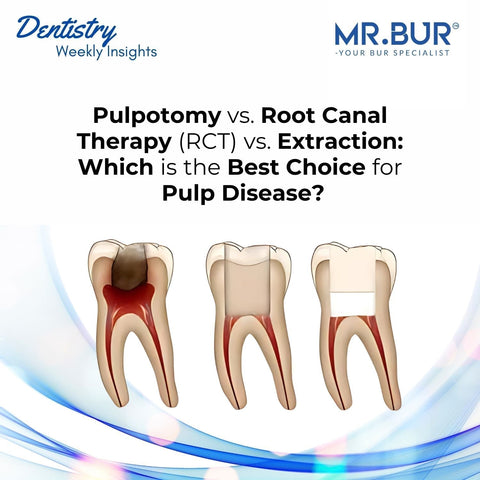
2. Pulp Necrosis (Death of the Pulp)

When pulp inflammation progresses without treatment, necrosis occurs due to a lack of blood supply, rendering the tooth non-vital.
Symptoms: The tooth may become asymptomatic initially but eventually lead to periapical pathology, causing pain, swelling, or abscess formation.
Treatment: Root canal therapy is necessary to remove the necrotic pulp, disinfect the canal, and prevent bacterial proliferation.
3. Periapical Abscess

A periapical abscess is a collection of pus at the root tip, resulting from bacterial invasion due to untreated pulp infection. It can lead to systemic complications if left unmanaged.
Symptoms: Throbbing pain, swelling, pus discharge, and potential fever.
Treatment:
-
Root canal treatment to remove infection.
-
Drainage if swelling is present.
-
Antibiotic therapy in cases of systemic involvement or spreading infection.
4. Internal Resorption

A rare condition where the pulp tissue undergoes resorption due to chronic inflammation, leading to progressive tooth weakening.
Symptoms: Asymptomatic in early stages but may present as a radiolucency within the tooth structure on radiographs.
Treatment: Root canal therapy is required to halt resorption and restore structural integrity.
Step-by-Step Treatment Approaches for Pulpal Diseases
1. Pulp Capping (For Reversible Pulpitis)

Step 1: Remove bacterial plaque and infected dentin
-
Ensure proper isolation using a rubber dam to prevent contamination.
-
Clean the tooth surface thoroughly.
Step 2: Access the cavity using appropriate burs
-
Use Mr. Bur round collar diamond bur in high-speed to remove unsupported enamel and open the cavity effectively.
Step 3: Remove infected dentin carefully
-
Use Mr. Bur round carbide bur in low-speed to remove affected dentin while preserving healthy structure.
Step 4: Disinfect and apply protective medicament
-
Disinfect the cavity with 2% chlorhexidine gluconate to eliminate remaining bacteria.
-
Apply calcium hydroxide or mineral trioxide aggregate (MTA) to protect the pulp and stimulate dentin formation.
Step 5: Seal the pulp protection layer
-
Apply glass ionomer cement to act as a liner.
-
Adjust occlusion using Mr. Bur pear shape diamond bur to ensure proper fit.
Step 6: Bonding process
-
Etch enamel margins with 37% phosphoric acid for 15 seconds (if using total-etch).
-
Apply a self-etching adhesive system to ensure optimal bonding.
Step 7: Restore with composite resin
-
Incrementally place composite restoration, curing each layer properly.
Step 8: Polish the restoration for a smooth finish
-
Use Mr. Bur Diamond Composite Polisher 6002 for final polishing and optimal aesthetics.
2. Root Canal Therapy (RCT) (For Irreversible Pulpitis, Necrosis, or Abscesses)

Step 1: Preoperative Assessment & Isolation
-
Conduct a clinical and radiographic examination to assess pulp vitality, canal morphology, and periapical status.
-
Ensure proper isolation using a rubber dam to maintain asepsis and prevent contamination.
Step 2: Access Cavity Preparation
-
Removes enamel and dentin to create initial access.
-
Provides straight-line entry into the pulp chamber.
-
Removes the roof of the pulp chamber for full exposure.
-
Assists in localizing canal orifices without overcutting.
Step 3: Canal Orifice Widening & Refinement
-
Smooths and refines the cavity walls for better canal access.
-
Creates a funnel-shaped access for better visibility.
-
Aids in precise and conservative shaping of the pulp chamber.
Step 4: Pulp Tissue Removal & Canal Cleaning
-
Use: Barbed Broaches or Endodontic Files
-
Removes necrotic pulp tissue and debrides canals.
-
Irrigate:
-
Use 2.5-5.25% Sodium Hypochlorite (NaOCl) for bacterial elimination.
-
Use EDTA (ethylenediaminetetraacetic acid) to remove the smear layer.
Step 5: Shaping the Root Canal System
-
Use: Nickel-Titanium Rotary or Hand Files
-
Enlarges and shapes canals for adequate cleaning and obturation.
-
Files should be used in a step-back or crown-down technique to ensure effective debridement.
Step 6: Final Disinfection & Drying
-
Perform final irrigation with NaOCl and saline rinse to remove debris.
-
Use sterile paper points to dry the canals completely.
Step 7: Obturation (Filling the Canal System)
-
Use: Gutta-Percha & Endodontic Sealer
-
Ensures complete canal sealing to prevent reinfection.
-
Can be placed via lateral condensation or warm vertical compaction technique.
Step 8: Coronal Seal & Restoration
-
Use: Glass Ionomer Cement (GIC) or Composite Base
-
Seals the access cavity to prevent microleakage.
-
Adjusts occlusion to ensure proper function and comfort.
-
If required: Crown Placement
-
A full-coverage crown is recommended for posterior teeth to protect against fractures.
Step 9: Final Polishing & Follow-Up
-
Smoothens the restoration for aesthetic and functional success.
-
Schedule periodic follow-ups with radiographs to monitor healing and long-term success.
3. Tooth Extraction (For Non-Restorable Teeth)

-
Step 1: Take radiographs to evaluate root structure and infection extent.
-
Step 2: Administer local anesthesia for pain control.
-
Step 3: Use elevators and forceps to carefully extract the tooth, minimizing trauma to surrounding tissues.
-
Step 4: Perform socket preservation (bone grafting) if future implant placement is considered.
-
Step 5: Provide post-extraction care instructions, including pain management and infection prevention.
4. Regenerative Endodontics (For Immature Necrotic Teeth in Young Patients)

-
Step 1: Remove necrotic tissue and disinfect the canal with gentle irrigation.
-
Step 2: Induce blood clot formation to create a scaffold for tissue regeneration.
-
Step 3: Apply growth factors or stem cell-based materials to stimulate pulp-like tissue regrowth.
-
Step 4: Seal the canal with biocompatible materials and monitor healing over time.
The dental pulp plays a pivotal role in maintaining tooth vitality, and its health is crucial for long-term dental function. Early diagnosis and intervention can prevent complications and preserve tooth structure. As advancements in endodontic therapy continue to evolve, regenerative and minimally invasive approaches are offering better outcomes for pulp preservation.
For dental professionals, staying informed about pulp pathologies and treatment modalities ensures improved patient care and long-term oral health success. If you're encountering complex pulp cases, consulting an endodontist may provide additional insight into the best treatment approach.
Diamond Burs, Carbide Burs, Surgical & Lab Use Burs, Endodontic burs, IPR Kit, Crown Cutting Kit, Gingivectomy Kit, Root Planning Kit, Orthodontic Kit, Composite Polishers, High Speed Burs, Low Speed Burs