Gingival recession is a prevalent concern in periodontal practice, often leading to root exposure, dentin hypersensitivity, and compromised aesthetics. As periodontists increasingly focus on soft tissue management, connective tissue grafting (CTG) has emerged as the gold standard for treating gingival recession. This article provides an in-depth review of the indications, technique, healing process, and success factors of CTG, offering insights that can enhance clinical outcomes and improve periodontal health worldwide.
1. Indications for Connective Tissue Grafting
CTG is primarily used in cases of Miller Class I and II gingival recession and is effective in:
- Covering exposed root surfaces to reduce sensitivity and decay risk.
- Increasing keratinized tissue width for improved periodontal stability.
- Enhancing esthetic outcomes in the anterior region.
- Preparing the periodontium for restorative or orthodontic procedures.
- Correcting thin biotype in patients at risk for further recession.
2. Surgical Technique of Connective Tissue Graft
A. Preoperative Assessment and Planning
Before performing CTG, a comprehensive periodontal evaluation is essential. The following factors should be assessed:
- Recession depth and width
- Periodontal attachment loss and pocket depth
- Keratinized tissue width
- Patient’s oral hygiene and compliance
- Occlusal trauma or parafunctional habits (e.g., bruxism)
Patient Preparation:
- Prophylactic scaling and root planing to remove bacterial deposits.
- Prescribing chlorhexidine mouthwash (0.12%) preoperatively.
- Local anesthesia using 2% lidocaine with epinephrine (1:100,000).
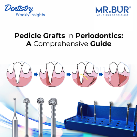
In cases where granulation tissue removal is required, Mr. Bur Degranulation Kit can be utilized. This kit provides specialized burs designed for efficient and precise debridement while preserving bone structure, ensuring a clean recipient site for grafting. The precision of these burs allows periodontists to effectively prepare the site while minimizing trauma to adjacent tissues, which is crucial for graft success.
B. Harvesting the Connective Tissue from the Palate
- A partial-thickness incision is made in the palatal donor site (between the first molar and canine).
- A subepithelial connective tissue graft (1.5-2 mm thick) is carefully dissected from beneath the epithelium.
- The donor site is sutured with resorbable 5-0 sutures or covered with a periodontal dressing.
C. Recipient Site Preparation
- The recipient area is debrided to remove inflamed tissue.
- A split-thickness flap is elevated to create a vascular bed for graft integration.
- The root surface is planed and, optionally, conditioned with EDTA or citric acid to enhance connective tissue attachment.
- Using Mr. Bur Degranulation Kit, the area is refined to ensure the removal of residual soft tissue debris while maintaining bone integrity, creating an optimal environment for graft placement.
D. Graft Placement and Suturing
- The harvested connective tissue is positioned over the exposed root.
- It is secured using resorbable sutures (6-0 or 7-0 Vicryl® /PTFE sutures) with interrupted or sling sutures.
- The flap is coronally advanced to fully or partially cover the graft.
- A periodontal dressing may be applied to protect the site.
3. Postoperative Healing and Patient Instructions
A. Healing Phases
- First 24-48 hours: Clot formation and initial tissue adhesion.
- Week 1: Epithelial proliferation; mild inflammation may be present.
- Week 2-4: Graft revascularization and integration.
- Week 6+: Maturation of new connective tissue and keratinized tissue formation.
B. Post-Operative Care Guidelines
- No brushing or flossing at the surgical site for 2 weeks.
- Chlorhexidine mouth rinse (0.12%) twice daily for 2-4 weeks.
- Soft diet for at least 7-10 days.
- NSAIDs (e.g., ibuprofen 400-600 mg every 6-8 hours) to manage pain.
- Avoid smoking, alcohol, and hard foods.
- Follow-up visits at 1 week, 1 month, and 3 months post-surgery.
4. Success Factors and Complications
A. Factors for a Successful Connective Tissue Graft
✅ Proper flap design and tension-free closure.
✅ Adequate blood supply to the graft.
✅ Sufficient donor tissue thickness (1.5-2 mm).
✅ Good plaque control and patient compliance.
✅ Stable suturing technique to prevent graft micromovement.
B. Potential Complications and Management
❌ Graft necrosis: Usually due to inadequate vascularization—consider re-evaluation and possible re-grafting.
❌ Palatal bleeding: Can be controlled with a hemostatic agent (e.g., Surgicel®) or compression.
❌ Excessive scarring or uneven gingival contour: May require minor recontouring after complete healing.
❌ Postoperative pain and sensitivity: Managed with topical anesthetics or desensitizing agents.
5. Long-Term Prognosis and Maintenance
Studies indicate that CTG has a 90%+ success rate, with graft stability maintained over 5–10 years, provided patients follow a strict oral hygiene regimen and attend regular maintenance visits. A keratinized tissue width of at least 2mm is considered essential for long-term stability.
Connective tissue grafting is a highly predictable and clinically effective technique for addressing gingival recession. By mastering proper harvesting and placement techniques, ensuring vascular integrity, and optimizing postoperative care, periodontists can achieve superior outcomes in both function and aesthetics.
By integrating modern tools like Mr. Bur Degranulation Kit, which enables efficient debridement without compromising bone integrity, clinicians can improve procedural efficiency and ensure a clean surgical field for successful graft integration. The precision of the kit facilitates superior results by minimizing tissue trauma and promoting optimal graft adaptation.
At MR.BUR UNITED KINGDOM, we pride ourselves on exceeding expectations and continually improving to meet the evolving needs of our customers.
Diamond Burs, Carbide Burs, Surgical & Lab Use Burs, Endodontic burs, IPR Kit, Crown Cutting Kit, Gingivectomy Kit, Root Planning Kit, Orthodontic Kit, Composite Polishers, High Speed Burs, Low Speed Burs


