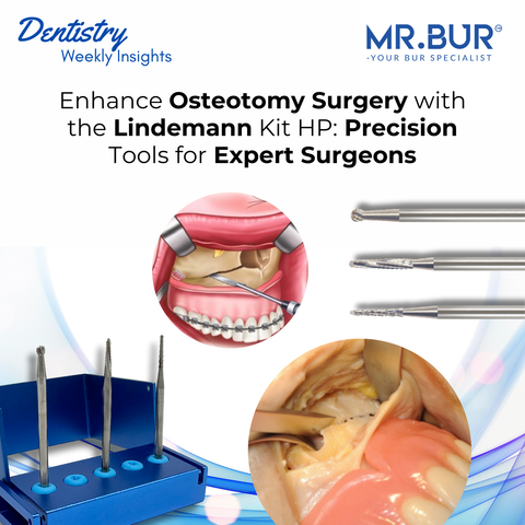
Sagittal Split Osteotomy (SSO) is widely regarded as the gold standard in orthognathic surgery for correcting mandibular malocclusion and improving facial symmetry. Achieving successful outcomes in sagittal split surgery requires precise bone cuts, controlled splitting, and minimizing trauma to the inferior alveolar nerve and surrounding tissues. Traditional cutting techniques often pose challenges such as bone splintering, uneven cuts, and nerve damage. This is where the Mr. Bur Lindemann Kit HP excels.
Mr. Bur Lindemann Kit HP, with its specialized surgical burs, is designed to provide precise bone cutting with minimal vibration and heat generation. Its ability to handle both cortical and cancellous bone with controlled efficiency makes it an invaluable tool for sagittal split surgery.
What is Sagittal Split Osteotomy (SSO)?

Sagittal Split Osteotomy is a surgical procedure used to reposition the mandible by cutting and splitting the bone along its sagittal plane. This procedure is commonly performed to correct:
-
Mandibular Retrognathia: When the lower jaw is positioned too far back.
-
Mandibular Prognathia: When the lower jaw protrudes too far forward.
-
Asymmetry: When one side of the jaw is longer or more prominent than the other.
-
Dental Occlusion Issues: When the upper and lower teeth do not align properly.
Key Techniques of Sagittal Split Osteotomy (SSO)
1. Incision and Flap Elevation

-
Make a horizontal incision along the anterior border of the mandibular ramus.
-
Extend the incision along the buccal surface toward the external oblique ridge.
-
Elevate a mucoperiosteal flap to expose the lateral and medial surfaces of the ramus.
-
Use a periosteal elevator to carefully elevate the soft tissue without damaging the underlying bone.
-
Ensure proper retraction of soft tissues and the inferior alveolar nerve.
2. Bone Cutting

Lateral Cortical Bone Cut
-
Use a surgical handpiece with Mr. Bur HP162 Lindemann Bur HP Bone Cutting to create a clean cut through the lateral cortical bone.
-
Start from the external oblique ridge and extend downward along the ramus.
-
Maintain a steady hand and consistent depth to avoid splintering.
Medial Cortical Bone Cut
-
Make a secondary cut along the medial surface of the ramus.
-
The medial cut should be parallel to the lateral cut but not extend completely through the bone.
-
Keep the inferior alveolar nerve protected at all times.
Inferior Border Cut
-
Extend the cut along the inferior border of the mandible to create a controlled splitting point.
-
Ensure the inferior border cut meets the lateral and medial cuts for a clean separation.
3. Bone Splitting

-
Insert an osteotome into the lateral cortical cut.
-
Gently tap the osteotome to initiate the splitting process.
-
Once the split is initiated, use bone spreaders to complete the separation.
-
Carefully maneuver the split segments to avoid bone fractures.
-
Confirm that the inferior alveolar nerve remains intact and undamaged.
4. Mandibular Repositioning and Fixation

-
Move the distal segment of the mandible into the desired position.
-
Ensure proper occlusal contact with the maxilla.
-
Check for symmetry and proper alignment.
-
Once the desired position is confirmed, use titanium plates and screws to secure the bone segments.
-
Typically, two or three plates are used to provide sufficient stability.
-
Confirm that the fixation is stable and there are no open contacts or occlusal discrepancies.
5. Smoothing and Refinement
-
Use Mr. Bur HP141E MOS Oral Surgical Round Carbide Bur HP to smooth any sharp edges or irregularities.
-
Ensure that the bone segments are flush and aligned.
-
Remove any bone spicules or rough surfaces to promote better healing.
6. Repositioning and Saturing
-
Reposition the mucoperiosteal flap over the surgical site.
-
Ensure the soft tissue is tension-free to avoid delayed healing.
-
Use resorbable sutures to close the incision.
-
Ensure proper adaptation of the mucosal tissue without excessive tension.
Common Complications and How to Avoid Them
Nerve Damage
-
Avoid direct contact with the inferior alveolar nerve during bone cutting.
-
Monitor nerve function during and after surgery.
Bone Fractures
-
Avoid excessive pressure during bone splitting.
-
Ensure even cuts and consistent bone thickness.
Post-Operative Infection
-
Maintain sterile technique throughout the procedure.
-
Prescribe antibiotics and monitor for signs of infection.
Sagittal Split Osteotomy remains the gold standard in orthognathic surgery due to its consistent success rate, versatility, and ability to improve both functional and esthetic outcomes. With advancements in surgical tools like the Mr. Bur Lindemann Kit HP, surgeons can achieve greater precision and control, resulting in faster healing and long-term stability.
Diamond Burs, Carbide Burs, Surgical & Lab Use Burs, Endodontic burs, IPR Kit, Crown Cutting Kit, Gingivectomy Kit, Root Planning Kit, Orthodontic Kit, Composite Polishers, High Speed Burs, Low Speed Burs